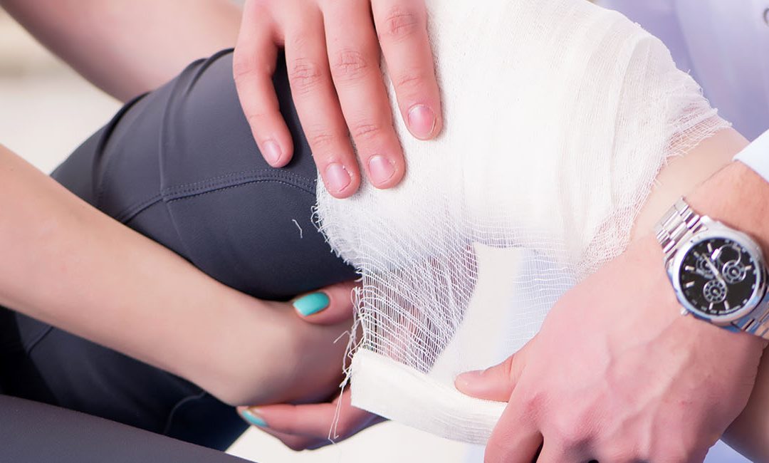
Distal femur is the lower end of the thigh bone which lies just above the knee joint and resembles an inverted funnel. The end of the bone is lined by a thick slippery substance called cartilage which allows it to slide across other bones that constitute the joint. It also helps in movement of the distal femur when the knee is bent. A crack or a break in this part of the bone is medically referred to as the Distal Femur Fracture of the Knee. In case the force causing the fracture is strong enough, it may also damage the kneecap. Such fractures are commonly observed in people aged above 50 years as their bones are relatively weaker. However, the younger populace is at an equal risk although the causative factors may vary. The injury can be classified as follows:
Closed Fracture – The skin is not ruptured
Open Fracture – The skin is cut open during the injury and a part of the bone may stick out
Comminuted Fracture – The injury causes the bone to shatter into multiple pieces
Transverse Fracture – The crack or breakage occurs straight across the bone These fractures may not only damage the femur but also affect the tendons and ligaments that surround it. The hamstring and the quadriceps muscles may tend to snap and shorten when the bone breaks.
Causes
- A fall from a height
- Vehicular accident
- Loss of bone strength and density as age increases
- Direct hit to the knee Sports injuries
Symptoms
- Severe pain
- Inability to stand or bear bodyweight
- Swelling
- The injured area may be tender when touched
- Visible deformity
- Change in gait as the leg may become crooked or shorter
- Diagnosis
- Clinical evaluation of the injured leg by an orthopedic doctor
- Evaluation of the mode of injury, patient’s medical records and the symptoms
- The blood supply to the nerves and their ability to feel sensations may be tested X-ray imaging, CT scan or MRI may be performed
Treatment
- If the bone is in a relatively stable condition and does not involve multiple fracture, it may be treated by using a cast or plaster
- The doctor may attach a traction pin to the bone and tie weights with a pulley. This may help to hold the bone in place.
- Surgery may be required in case of an open fracture to avoid infection. The broken bone may be put in place using external fixator devices (screws and pins) which are fixed on a frame attached to the leg.
- Internal fixation may also be done by making surgical incisions. A metal rod may be inserted to keep the femur in place
- Severely damaged bone may need a bone graft which involves the extraction of a piece of bone from the pelvis.
- Artificial bone fillers or allograft may be used in some cases
- Knee replacements may be recommended for elderly patients
- Physical therapy may be required in the post-operative phase to aid healing and restore flexibility as well as strength in the leg


